Empower Your Health

Musculoskeletal Scans

All pictures 48 of 55 1
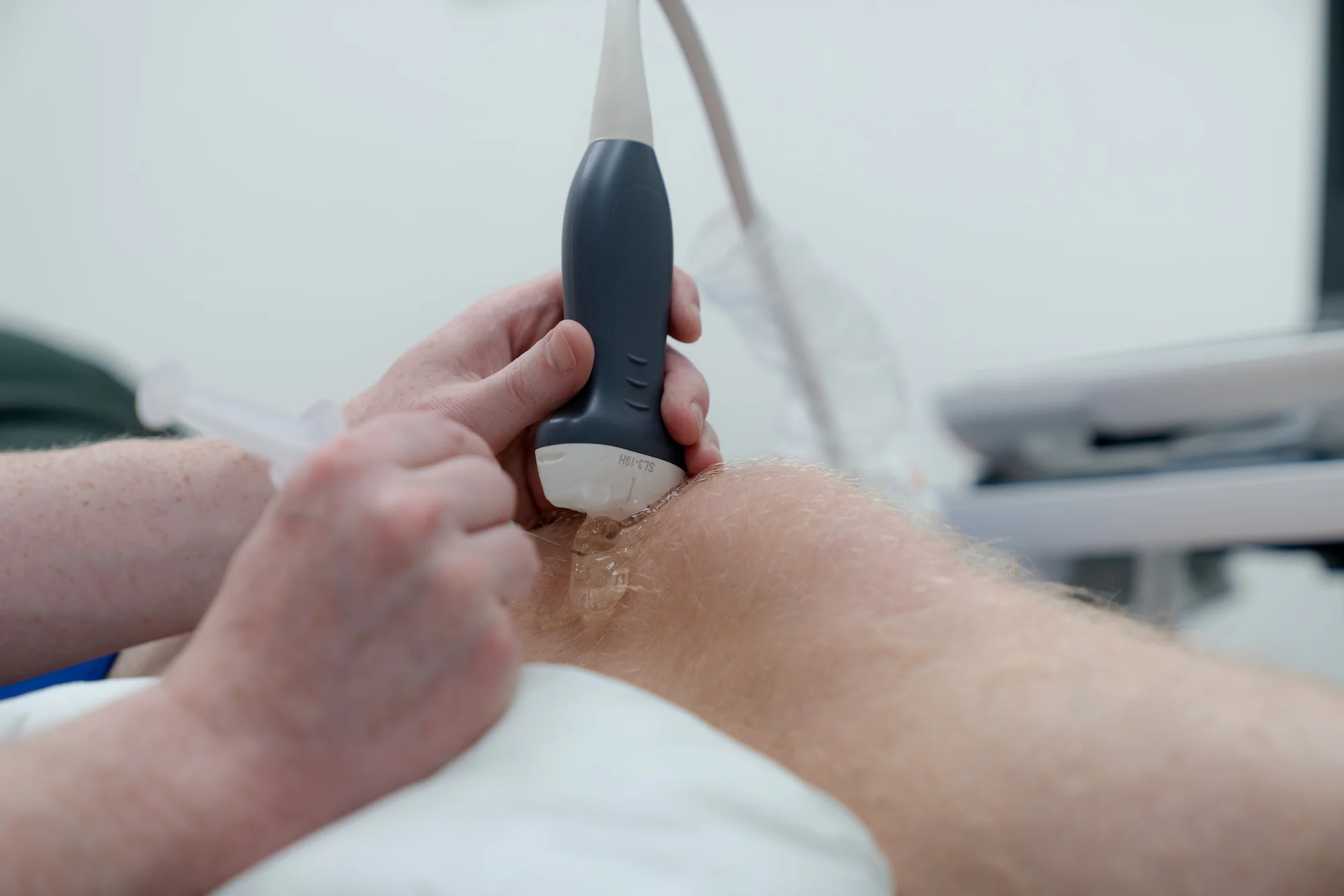
At Ultrasound Scans Motherwell, we specialize in providing high-quality musculoskeletal ultrasound scans to help diagnose and manage a wide range of joint, tendon, ligament, and muscle conditions. Our team of experienced professionals uses the latest ultrasound technology to provide accurate, real-time imaging, enabling us to identify the root cause of your pain or discomfort. Whether you’re experiencing sports injuries, chronic pain, or any musculoskeletal issues, we are here to offer a non-invasive, effective solution for accurate diagnosis and tailored treatment plans.
 
Our clinic is equipped with state-of-the-art ultrasound machines, ensuring precise imaging for even the most complex musculoskeletal conditions. Musculoskeletal ultrasound is an invaluable tool for detecting soft tissue injuries, inflammation, tears, and other abnormalities in the muscles and joints. During the scan, our skilled sonographers will guide you through the process, explaining each step and providing clear insight into your results, so you feel confident in your diagnosis and next steps.
 
At Ultrasound Scans Motherwell, we understand that musculoskeletal conditions can be disruptive to your daily life, and we are committed to providing a fast, reliable, and stress-free experience. We offer flexible appointment scheduling and ensure that each scan is performed in a comfortable, professional setting. With fast, accurate results, you can be assured that you’ll receive the highest level of care to help manage and treat your musculoskeletal health. Contact us today to schedule your musculoskeletal ultrasound scan and take the first step toward recovery.
 

Ultrasound is commonly used to assess shoulder pain and dysfunction, particularly in evaluating soft tissue injuries and joint abnormalities. Below are some of the common anatomical structures and diagnoses that can be seen on ultrasound.

  • Anatomy: Rotator cuff tendons (supraspinatus, infraspinatus, subscapularis, teres minor), deltoid, biceps tendon (long and short head), acromioclavicular joint, bursae.
  • Common Diagnoses: Rotator cuff tears/tendinopathy, bursitis (subacromial/subdeltoid), biceps tendinopathy/tear, calcific tendinopathy, impingement syndrome, arthritis.

Ultrasound is useful in detecting soft tissue and vascular conditions of the upper arm, particularly injuries to the biceps and triceps muscles. Below are some of the common anatomical structures and diagnoses that can be seen on ultrasound.

  • Anatomy: Biceps brachii, triceps brachii, brachialis, brachial artery, nerves (musculocutaneous, radial, median, ulnar).
  • Common Diagnoses: Biceps/triceps tendon tears or tendinopathy, muscle strains, myositis ossificans, hematomas, nerve entrapment.

Ultrasound provides real-time imaging of tendons, ligaments, and nerves in the elbow and forearm, aiding in the diagnosis of common overuse injuries. Below are some of the common anatomical structures and diagnoses that can be seen on ultrasound.

  • Anatomy: Common extensor tendon (lateral epicondyle), common flexor tendon (medial epicondyle), ulnar collateral ligament, radial collateral ligament, olecranon bursa, median/radial/ulnar nerves.
  • Common Diagnoses: Lateral epicondylitis (tennis elbow), medial epicondylitis (golfer’s elbow), bursitis (olecranon), ligament injuries (UCL/RCL tears), nerve entrapments (cubital tunnel syndrome, radial tunnel syndrome), muscle strains.

Ultrasound is a valuable tool for assessing tendon injuries, nerve entrapment, and inflammatory conditions affecting the wrist, hand, and fingers. Below are some of the common anatomical structures and diagnoses that can be seen on ultrasound.

  • Anatomy: Carpal tunnel (median nerve, flexor tendons), extensor tendons, flexor tendons, triangular fibrocartilage complex (TFCC), intrinsic muscles, ligaments.
  • Common Diagnoses: Carpal tunnel syndrome, De Quervain’s tenosynovitis, tendon ruptures or inflammation, TFCC tears, ganglion cysts, arthritis, trigger finger, ligament sprains/tears

Hip ultrasound is frequently used to evaluate tendon injuries, bursitis, and joint abnormalities, especially in active individuals and athletes. Below are some of the common anatomical structures and diagnoses that can be seen on ultrasound.

  • Anatomy: Hip joint, femoral head/neck, acetabulum, iliopsoas tendon, gluteal tendons, hamstring origin, trochanteric bursa.
  • Common Diagnoses: Hip joint effusion, hip osteoarthritis, iliopsoas tendinopathy, femoroacetabular impingement (FAI), trochanteric bursitis, gluteal tendinopathy, hamstring tendinopathy/tear, snapping hip syndrome.

Ultrasound is highly effective in assessing soft tissue injuries of the thigh, particularly muscle tears and hematomas. Below are some of the common anatomical structures and diagnoses that can be seen on ultrasound.

  • Anatomy: Quadriceps (rectus femoris, vastus lateralis, vastus medialis, vastus intermedius), hamstrings (biceps femoris, semitendinosus, semimembranosus), adductors, sciatic nerve, femoral artery/vein.
  • Common Diagnoses: Muscle strains/tears (hamstrings/quadriceps), hematomas, myositis ossificans, nerve entrapment, deep vein thrombosis (DVT).

Ultrasound plays a crucial role in evaluating knee injuries, particularly those involving tendons, ligaments, and bursae. Below are some of the common anatomical structures and diagnoses that can be seen on ultrasound.

  • Anatomy: Quadriceps tendon, patellar tendon, medial/lateral collateral ligaments, menisci (medial/lateral), joint capsule, bursae, popliteal fossa.
  • Common Diagnoses: Patellar tendinopathy (jumper’s knee), quadriceps tendinopathy, ligament injuries (MCL/LCL sprains), meniscal cysts, Baker’s cyst, bursitis, joint effusion, tendon tears, avulsion fractures.

Ultrasound is an effective modality for assessing muscle injuries, tendon pathology, and vascular conditions in the lower leg. Below are some of the common anatomical structures and diagnoses that can be seen on ultrasound.

  • Anatomy: Gastrocnemius, soleus, tibialis anterior, peroneal muscles, Achilles tendon, deep posterior compartment, tibial and peroneal nerves, blood vessels.
  • Common Diagnoses: Muscle strains (calf tears), Achilles tendinopathy/rupture, shin splints (medial tibial stress syndrome), compartment syndrome, DVT, stress fractures.

Ultrasound is widely used to diagnose soft tissue injuries, tendon pathologies, and ligament sprains affecting the foot and ankle. Below are some of the common anatomical structures and diagnoses that can be seen on ultrasound.

  • Anatomy: Achilles tendon, peroneal tendons, plantar fascia, extensor/flexor tendons, tibialis posterior, ankle ligaments (ATFL, CFL, PTFL), joints (subtalar, midfoot, MTP joints).
  • Common Diagnoses: Achilles tendinopathy/rupture, peroneal tendinopathy, plantar fasciitis, ligament sprains/tears (ATFL sprain), tarsal tunnel syndrome, Morton’s neuroma, stress fractures, arthritis.